中国科学院微生物研究所,中国微生物学会,中国菌物学会
文章信息
- Ruihua Lü, Panpan Zhu, Maode Yu, Changying Liu, Aichun Zhao. 2019
- 吕蕊花, 朱攀攀, 余茂德, 刘长英, 赵爱春. 2019
- Conidial formation and pathogenicity of Ciboria shiraiana
- 桑椹肥大性菌核病菌(Ciboria shiraiana)分生孢子的形成和致病性
- Acta Microbiologica Sinica, 59(12): 2367-2377
- 微生物学报, 59(12): 2367-2377
-
文章历史
- 收稿日期:2019-01-29
- 修回日期:2019-04-10
- 网络出版日期:2019-04-22
2. College of Medical Technology, Shaanxi University of Chinese Medicine, Xianyang 712046, Shaanxi Province, China
2. 陕西中医药大学 医学技术学院, 陕西 咸阳 712046
Mulberries are juicy sweet-tasting fruits rich in essential amino acids, vitamins, flavones and anthocyanins, and are listed as a medicinal food. However, a devastating disease, 'mulberry fruit sclerotiniosis' can occur during mulberry cultivation in the USA[1-3] and other countries worldwide, and is especially severe in rainy and humid regions, such as Sichuan, Chongqing, Zhejiang, Jiangsu, and Guangdong Provinces in China, where incidence rates have reached 30%-90%, with this rate increasing each year[4]. The apothecia of the mulberry fruit sclerotiniosis pathogens germinate in late February from overwintered sclerotia, which remain wrapped inside the drupelets in the former year. Ascospores that are released from apothecia serve as important primary inoculum sources of mulberry fruit[1, 5].
The pathogens of mulberry fruit sclerotiniosis include Ciboria shiraiana, Mitrula shiraiana, and Ciboria carunculoides, with the former having the greatest apothecial germination rate, resulting in the most serious damage during mulberry cultivation. C. shiraiana (Ascomycotina, Sclerotiniaceae, Ciboria) is the pathogen of hypertrophy sorosis scleroteniosis, which is known in other countries as popcorn disease, swollen-fruit disease, or shrunken-fruit disease[5]. The features of the pathogen include atmospheric dissemination and a high mutation rate. Despite numerous efforts to control the disease, currently the most effective method relies on the use of fungicides[6]. Recently, other measures were explored, such as narrow windrow burning of canola residue[7], biocontrol agents like Bacillus thuringiensis C25[8], cuminic acid isolated from the seed of Cuminum cyminum L.[9], and native lipopeptide-producer Bacillus strains[10].
Conidia are a kind of asexual diaspore produced during the asexual reproductive stage in Ascomycetes and Deuteromycetes. In Sclerotiniaceae, only a minority of genera, such as Botryotinia and Monidinia[11], produce conidia during their life cycles. Conidia are considered an infection source, but the infectivity rates are much lower than that of ascospores. As in grape, the conidia of Botrytis cinerea infect mainly the flower receptacles and, to a lesser extent, the stigma and styles[12]. Conidial function may be related to the formation of ascogonia, which are beneficial for the germination of sclerotia into apothecia. Conidial formation can be stimulated by specific wavelengths of light, and near UV is generally used to induce sporulation in cultures[13-14]. Relative humidity (RH) is also a crucial environmental factor for conidial germination. For example, conidia of B. cinerea require a RH value above 93% to infect rose petals[15].
In Sclerotiniaceae, AC, which is activated by three genes encoding Gα subunits of heterotrimeric G-proteins, starts the synthesis of cAMP, and the increased cAMP level intracellularly activates a variety of factors, including effector molecules, leading to a variety of PKA-regulated physiological functions[16], including sclerotial development and conidial formation in S. sclerotiorum[17]. AC, which catalyzes cAMP formation, is usually considered a key factor for conidial formation in Sclerotiniaceae, as an AC-deficient mutant strain was shown to have altered hyphal branching pattern, more abundant microconidia, abnormal sclerotia and a lower virulence level[16]. An exogenously supplied concentration of more than 5 mol/L cAMP completely inhibits sclerotial formation in Sclerotinia sclerotiorum, indicating that cAMP plays a significant role in the early transition from mycelial growth to sclerotial development[18].
Conidiophores and conidia of C. shiraiana were first shown as graph patterns, with hyphae spread on the perianth tissues and conidiophores produced in the ovaries[2], but the conditions for optimum production and pathogenicity of conidia in the field and laboratory have not been ascertained until now. The objectives of this study were to determine whether conidia from diseased mulberry fruit could infect healthy fruit, and to establish a feasible and operable method to produce conidia through artificial culturing. Furthermore, the influence of cAMP on conidial formation was investigated at different stages of sclerotial development. These results will increase our understanding of the environmental conditions required for mulberry fruit infection, as well as the complete infection cycle and the conidial formation pathway in C. shiraiana.
1 Materials and methods 1.1 Formation and pathogenicity of conidia in diseased mulberry fruitTo prevent natural infections by ascospores of C. shiraiana, mulberry fruits of "No. 40 Jialing" (Morus atropurpurea Roxb.) were bagged before winter buds had germinated in the mulberry orchard of Southwest University, Chongqing, China in early February 2013 and 2016. To obtain mulberry fruits with hypertrophy sorosis scleroteniosis, in early March when mulberry fruits were in bloom, more than 1×10⁴ bagged mulberry fruits previously were inoculated with ascospore of C. shiraiana at about 8:00 a.m, which were induced in our laboratory according to the method of Wang et al.[19], and then wrapped with plastic film to stay moist. Conidiophores and conidia produced in diseased mulberry fruit were observed with the aid of a biomicroscope (Olympus America, Inc., Center Valley, PA, USA). The surface structures of conidia were observed using a scanning electron microscope (SEM; Nikon, Tokyo Japan), after preparing samples following the methods of Andreas et al.[20].
The inoculated mulberry fruits with ascospores showed typical symptoms of hypertrophy sorosis scleroteniosis, which we divided into five stages (Figure 1-A). The stage that diseased mulberry fruits could produce the maximum number of conidia were surface sterilized, ground into discrete conidia and filtered through sterilized lens paper, and then, the conidial suspension was diluted with sterile water to a concentration of 1×10⁶/mL. Every 2 d, about 1×10³ bagged mulberry fruits previously were inoculated with the conidial suspension, with sterile water as the control, when mulberry female flowers were in full blossom, from late March to early April. Because the maturation period of mulberry fruit can last 20-30 d, depending on the mulberry variety, about 1×10³ freshly diseased mulberry fruits of each stage were tied to healthy fruits using ropes to observe whether the latter became infected. To compare the pathogenicity levels between conidia and ascospores of C. shiraiana, mulberry fruits were inoculated using ascospores (1×10⁶ /mL) as above. When mulberry fruit was ripe, counted the number of diseased mulberry fruits and the total number of mulberry fruits, and the ratio was disease incidence. Conidia were quantified using a hemocytometer after being randomly collected from 10 diseased mulberry fruits of different stages using the following procedure: diseased mulberry fruits were mixed using a glass homogenizer after adding 5 mL water. Then, 1 μL of the liquid was placed on the hemocytometer, and the number of conidia in each diseased mulberry fruit was calculated. Experiments were repeated three times. Temperatures and RH levels were recorded daily from March 1 to April 30, 2013. These operations were repeated in 2014 and 2015.
|
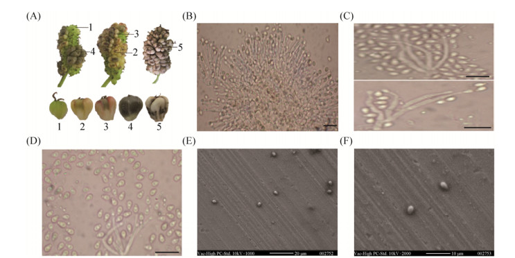
| Figure 1 Conidiophores and conidia in diseased mulberry fruit having hypertrophy sorosis scleroteniosis. (A) Mulberry fruit 15 d after being inoculated with conidia of C. shiraiana; the five disease stages are indicated; (B) Conidiophore produced by the mycelia; (C) and (D) Conidia separated from the top of the conidiophore; (E) and (F) Conidial morphological features under SEM. Bar=10 μm. |
1.2 Artificial conditions required for C. shiraiana conidia production
To induce conidial formation, hyphae of C. shiraiana (strain number: CCTCC AF 2014019 WCCQ01 Ciboria shiraiana), which were isolated and preserved on PDA medium in previous work[21], were inoculated into different media, PDA, PDA without sugar (PA) and potato dextrose broth (PDB), for PDB media with or without shaking at 200 r/min. As well as potato slices (PS; Φ=80 mm, h=2 mm), sweet potato slices (SPS; Φ=80 mm, h=2 mm), purple sweet potato slices (PSPS; Φ=50 mm, h=2 mm), carrot slices (CS; 1.5 cm×6 cm, h=2 cm), mulberry fruit (MF), and 1.2% water agar containing 0.2 g mulberry fruit powder (WAM). All of the potatoes, sweet potatoes, purple sweet potatoes, and carrots were purchased from a supermarket in China. The medium that induced the highest conidia yield was termed the inducing medium (IM). To determine whether light affected conidial formation, all of the media were placed in incubators set at either 24 h darkness or 12 h light/12 h dark, using either an incandescent lamp, ultraviolet light, or black fluorescence as the light source. In addition, a piece of nylon membrane was placed on the IM to determine whether nutrient substances affected conidial formation. All of the media were sterilized by autoclaving for 20 min at 121 ℃. The testing of every medium was repeated three times, with three replicates each.
1.3 Effects of temperature and RH on conidial formation under artificial conditionsTo determine the influence of temperature and RH on conidial formation, dishes containing IM inoculated with hyphae were placed at temperatures ranging from 10 ℃ to 40 ℃, at intervals of 5 ℃, and RH values of 40%, 50%, 60%, 70%, 80%, 90%, and 95% were used in light incubators. The temperature that produced the maximum number of conidia was used for the RH determinations. Conidia were obtained by washing the IM surface every day with 5 mL diluted water, and the number of conidia produced by every square centimeter of hyphae was quantified in three independent samples using a hemocytometer under a microscope.
1.4 Germination rate and infectivity of conidia under artificial conditionsThe germination rate of conidia was determined every 2 h under a microscope after culturing in a 25 ℃ incubator in water. Conidia were judged to have germinated when the germ tube length was at least equal to the diameter of the conidia. To survey the infectivity of conidia, conidia were collected as described above. In mid-March, a 1×10⁵/mL suspension of conidia was used to inoculate mulberry fruits that were bagged in February when the mulberry female flowers started to blossom. Symptoms of infected mulberry fruits were observed every 2 d.
1.5 The cAMP pathway in conidial formationTo study the influence of the cAMP pathway on conidial formation, different concentrations of cAMP (2, 5 and 10 mol/L)[22] were used independently as supplements every 24 h from 0 to 8 d on IM, using the same sucrose solution concentration as the control. Each concentration cAMP repeated three times with 8 dishes, and results were observed every 24 h under microscope. Hyphae and sclerotia from diseased mulberry fruit (stage 1 to 5), which were collected from PDA and IM, respectively, were used as materials. Total RNAs were extracted following the RNAiso Plus instructions (TaKaRa), treated with DNase and subjected to first-strand DNA synthesis using M-MLV reverse transcriptase. We determined the relative expression levels of AC and PKA in the cAMP pathway, using β-tubulin as an internal reference (Table 1). PCR was performed using SYBR Premix Ex TaqTM Ⅱ and the StepOne PlusTM Real-Time PCR System (Applied Biosystems), with the following reaction program: 95 ℃ for 30 s, and 40 cycles at 95 ℃ for 5 s, 60 ℃ for 13 s, and 95 ℃ for 15 s, followed by 60 ℃ for 1 min and 95 ℃ for 15 s. The total volume of each PCR mixture was 20 μL and consisted of 10 μL of SYBR Premix Ex Taq Ⅱ (2×), 0.4 μL of ROX Reference Dye (50×), 0.8 μL of each primer, 2 μL of cDNA, and 6 μL of double-distilled H2O. All of the qRT-PCR reactions were performed at least in triplicate. The gene specific primers are listed in Table 1, and the relative quantifications of the gene transcripts were calculated using the method of 2-ΔΔCT as described by Livak and Schmittgen[23].
| Primer name | Primer sequence (5′→3′) |
| AC-F | AGGATATTCAAAGCGGATGG |
| AC-R | CGAGACTGTCAGCCTGGTAA |
| PKA-F | ATCTTGCGCCAGAGGTTATT |
| PKA-R | AATATACCCAAAGCCCACCA |
| β-tubulin-F | TTGGATTTGCTCCTTTGACCAG |
| β-tubulin-R | AGCGGCCATCATGTTCTTAGG |
2 Results 2.1 Formation and pathogenicity of conidia in diseased mulberry fruit
Conidia appeared in diseased mulberry fruit 15 d after being inoculated with discrete conidia of C. shiraiana, and the resulting symptoms were the same as those infected with ascospores after 12 to 15 d. We divided the symptoms into five stages (Figure 1-A); during stage 1 and stage 2, there were no conidiophores or conidia on the diseased mulberry fruit. Conidiophores, which formed from specialized hyphae, were observed in stage 3. Each conidiophore appeared like a stick, with a thick bottom and a thin top, and they were 0.8-2.1× 7.6-25.2 μm in size (Figure 1-B). In stage 4, clusters of conidiophores segregated from hyphae, and a large number of conidia separated from the top of the conidiophore (Figure 1-C and D). In stage 5, mulberry fruit, hyphae, conidia, and conidiophores, which were wrapped in the peel of every drupelet, constituted a flinty sclerotium, with an outer black rind and white inner part. The swollen white fruit formed black sclerotia in the end.
Conidia appeared oval, shaped like grape seeds, and were colorless, well-stacked, and transparent. They measured (1.1-1.9) μm×(2.43-3.78)μm under SEM (Figure 1-E, F). In stage 3, 9×10⁴ conidia appeared on every diseased mulberry fruit compared with 8×10⁸ conidia in stage 4. The germination rate was very low for conidia detached from mulberry fruit, which formed short-lived propagules.
The rope-tying experiment showed that contact with the diseased mulberry fruits resulted in a 79.3% disease incidence in healthy mulberry fruits, while 37% and 90% disease incidences were found with the conidia and ascospore suspensions, respectively. Thus, ascospore play the greatest role in pathogenicity.
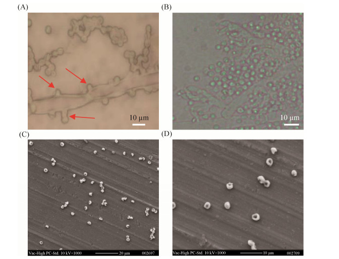
2.2 Artificial conditions required for C. shiraiana conidia productionConidiophores and conidia only appeared on fluffy mycelia after 8 d of incubation when using autoclaved PS as the medium; therefore, PS was used as the IM. Conidiophores and conidia were not observed on the fluffy hyphae grown on other media. Light conditions had no effect on the formation of conidia on the IM, but a limited number of conidia were observed when hyphae on IM were exposed to ultraviolet light or black fluorescence. Hyphae inoculated on a nylon membrane above the IM did not produce conidiophores or conidia. Conidiophores and conidia began to differentiate from single mycelia after 8 d of incubation at room temperature (Figure 2-A). From the 9th d, clusters of conidiophores appeared, measuring (0.8-2.1) μm × (0.6-2.8) μm. Then, after 10 d of culturing, masses of conidia fractured from the top of the conidiophores. The conidia formed on the IM were circular and colorless, and had particles in the middle when observed under a microscope (Figure 2-B). Further observations revealed that the shape was round, ranging from 2.6 to 3.6 μm, and the observed particles were sunken in the middle when observed under SEM (Figure 2-C, D).
|
| Figure 2 Conidiophores and conidia of C. shiraiana produced on inducing media. A: conidiophores and conidia started to differentiate from single mycelia 8 d after incubation (marked with arrows); B: conidia were released from the top of the conidiophore after 10 d; C and D: conidial morphological features under SEM. |
2.3 Suitable temperature and RH for conidial formation under artificial conditions
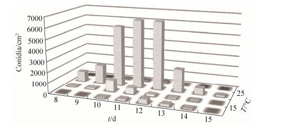
C. shiraiana began to produce conidia on IM after 8 d of culturing at 20 and 25℃. Mycelia did not grow at temperatures greater than 35℃, and no conidia formed at temperatures greater than 35℃ or less than 10 ℃. Conidia were produced from 8 to 15 d consistently after inoculation on IM when incubated at 15-30 ℃. Maximum production occurred at 25 ℃, ranging from 1.77×10³ to 6.37×10³ conidia/cm2 (Figure 3). The temperatures usually range from 14 ℃ to 29 ℃ from early March to late April in Chongqing, China, which is highly suitable for conidiophore and conidial formation.
|
| Figure 3 Effect of temperature on the conidial formation of Ciboria shiraiana (conidia/cm2 media) every day in culture. |
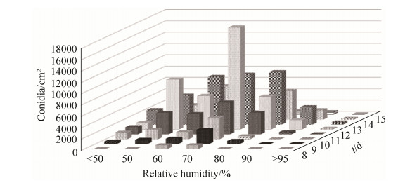
The production and formation rates of conidia were affected significantly by RH, with the earliest production occurring at 60% and 70% RH. C. shiraiana produced abundant conidia when the RH was between 50% and 80%, and there was a lower conidial yield beyond these RH levels at 25 ℃ (Figure 4). It was beneficial for aerial mycelia of C. shiraiana to grow and reproduce at a greater than 90% RH.
|
| Figure 4 Effect of relative humidity on the conidial formation of Ciboria shiraiana (conidia/cm2 media) every day in culture. |
2.4 Germination rate and infectivity of conidia under artificial conditions
The germination rate was very low at 25 ℃, only 3.4%, when incubated in water for 12 h after conidia being washed from the IM. Conidia started to germinate after 6 h of culture, and developed branching germ tubes after 12 h. The conidia produced on IM did not appear to be pathogenic to mulberry fruits because no typical symptoms were observed.
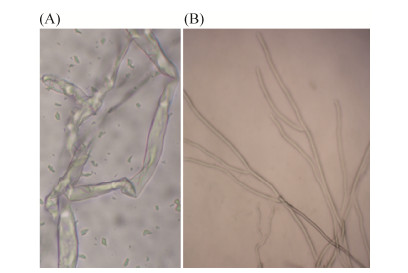
2.5 The cAMP pathway in conidial formationcAMP was used as an exogenous substrate added to the IM, and a cAMP concentration of 5 mol/L or greater inhibited conidial formation from the 1st to 5th day, with mycelia being morphologically abnormal (Figure 5). Any concentration of cAMPs blocked conidial formation and affected mycelial morphology after the 6th day. However, cAMPs, as a supplement to the IM, did not affect sclerotial formation. The cAMP produced by adenylyl cyclase serves as an important regulatory signal by activating downstream protein kinases (eg. cAMP-dependent protein kinase A, PKA) or transcription factors[24]. In the present work, cAMP, as an exogenous supplement to the IM, inhibited conidial formation, possibly by interfering with a subunit of PKA, which inhibited its regulatory function.
|
| Figure 5 Effect of cAMP as exogenous on hypha of C. shiraiana. A: 5 mol/L cAMP was used as supplements on hypha; B: Control. |
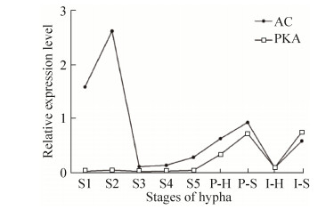
A qPCR analysis showed that the relative transcript levels of AC increased in stage 2, a 2.8-fold in diseased mulberry fruit, but sharply declined in stages 3 and 4. There was no expression of PKA from stages 1 to 5. Both AC and PKA in the sclerotia that were produced on PDA and IM, respectively, had greater expression levels than in hyphae produced on PDA and IM, respectively, because sclerotia formation precedes conidiation (Figure 6). Both AC and PKA were lower in hyphae produced on IM than in those produced on PDA, further indicating that the cAMP pathway negatively regulates conidial formation.
|
| Figure 6 qPCR analysis of the relative levels of AC and PKA expressed transcripts at different developmental stages. Stage S1-S5: Diseased mulberry fruit having hypertroophy sorosis scleroteniosis, shown in Figure 1; P-H and P-S indicate hyphae and sclerotinia produced on PDA, respectively; I-H and I-S indicate hyphae and sclerotinia produced on inducing media, respectively. |
3 Conclusion
In this report, we used M. atropurpurea Roxb. "No. 40 Jialing" as a research material; however, we observed other mulberry cultivated varieties, such as M. atropurpurea Roxb. "Da10", Morus alba L. "Zhenzhubai", M. alba "No. 1 Hongguo", M. alba "No.2 Hongguo" and M. atropurpurea Roxb. "No. 30 Jialing", to determine whether conidiophores and conidia existed in diseased mulberry fruit having hypertrophy sorosis scleroteniosis. In all of the varieties in which conidiophores and conidia could be observed, the conidial formation process was as observed in M. atropurpurea Roxb. "No. 40 Jialing". This indicated that conidiophores and conidia were prevalent in diseased mulberry fruits having hypertrophy sorosis scleroteniosis.
Only autoclaved PS induced conidiophores and conidia after 8 d of incubation. We suspect that the conidiophore and conidial production of C. shiraiana depends on the existence of a substrata, because hyphae could not produce conidia when inoculated on a nylon membrane above the IM. In Sclerotiniaceae, the sclerotia could be divided into sclerotial stroma and substratal stroma. The development of the sclerotial stroma, a melanized hyphal aggregate, is the common characteristic of all members of the Sclerotiniaceae[25]. However, the substratal stroma is an irregular structure consisting of penetrating tissue mycelia and host tissue[26]. For diseased mulberry fruit having hypertrophy sorosis scleroteniosis, the pathogen infects female mulberry fruit when they are in the early flowering or flowering period. After mycelia propagate in the pericarp of mulberry fruit, flinty sclerotia can be seen; therefore, the stroma of C. shiraiana in mulberry fruit should be termed substratal stroma. Thus, flat agar surfaces, such as those of PDA, PA and WAM, might be unsuitable for conidial formation. Conidia were not observed on mulberry fruit, probably because certain nutrients were destroyed after autoclaving. Additionally, media with high sugar contents are suitable for the growth of mycelia; however, the sugar contents were much greater in SPS and PSPS than in PS, which may have prevented conidial formation. Therefore, the production of conidiophores and conidia may be based on the substrate and the presence of a moderate carbon source. The function of conidia in sclerotia formation is not clear at present, and we hypothesize that the function is related to the development of ascogonia, which are beneficial for the germination of sclerotia into apothecia.
In this article, we used diseased mulberry fruits and dispersed conidia to infect healthy mulberry fruits, causing secondary infections. There are 2-8 mulberry female flowers in a winter bud, and the florescence of mulberry flowers occurs from early to late March. Therefore, conidia from one diseased mulberry fruit can infect healthy winter buds through physical contact during this phase. From our observations, it requires 12-15 d for typical symptoms to appear after healthy mulberry fruits are inoculated with conidia. In late March, newly diseased mulberry fruits are visible, making it possible for the produced conidia to reinfect healthy fruits. At present, it is better to choose mulberry varieties having a certain clearance between branches to avoid conidial-based infections.
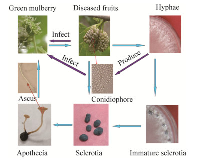
Our surveys further complement the knowledge of the C. shiraiana infection cycle. Conidia can be produced in diseased mulberry fruit as a source of secondary infections, or they can be induced on IM (Figure 7). Previously, in 2013 and 2014, we used hyphae of C. shiraiana cultured on PDA media as an infection source to infect mulberry fruits, but the hyphae did not cause an infection. Because both ascospores and conidia can cause infection, the removal of diseased mulberry fruit could be an effective method to control the spread of hypertrophy sorosis scleroteniosis.
|
| Figure 7 Complete infection cycle of Ciboria shiraiana. |
In China, the incidence of hypertrophy sorosis scleroteniosis in the Yangtze River region is significantly higher than that in the Yellow River region[4]. As the RH is usually between 60% and 80% from February to April near the Yangtze River and between 25% and 70% near the Yellow River. Besides, the temperature is more suitable for conidial formation in Yangtze River (10-25 ℃) from early to late March.
Adenylyl cyclase converts ATP to form cAMP and pyrophosphate, providing the primary source of intracellular cAMP. AC was expressed at a greater level in stage 2, while it sharply decreased in stages 3 and 4, which could result in the production of conidiophores and conidia. The cAMP pathway negatively regulated conidial formation, showing a strong inhibitory effect on AC, ΔPdac1 produces more conidia than wild-type progenitors, indicating that Pdac1 negatively regulates conidial formation in P. digitatum[27]. The expression levels of AC and PKA were significantly lower in stage 5 than those in sclerotia produced on PDA and IM, probably because conidia were contained in the sclerotia of the diseased mulberry fruit. The relative levels of AC and PKA expression were lower in hyphae produced on IM compared with those produced on PDA; however, the same levels of AC and PKA were expressed in sclerotia produced on PDA and IM, further indicating the negative regulatory function.
| [1] | Siegler EA, Jenkins AE. A new sclerotinia on mulberry. Science, 1922, 55(1422): 353-354. DOI:10.1126/science.55.1422.353 |
| [2] | Gray E, Gray RE. Observations on popcorn disease of mulberry in south central Kentucky. Castanea, 1987, 52(1): 47-51. |
| [3] | Hong SK, Kim WG, Sung GB, Nam SH. Identification and distribution of two fungal species causing sclerotial disease on mulberry fruits in Korea. Mycobiology, 2007, 35(2): 87-90. DOI:10.4489/MYCO.2007.35.2.087 |
| [4] | 黄尔田, 田立道, 肖练章, 周水良. 实用桑树保护学. 成都: 四川科学技术出版社, 1992. |
| [5] | Whetzel HH, Wolf FA. The cup fungus, Ciboria carunculoides, pathogenic on mulberry fruits. Mycologia, 1945, 37(4): 476-491. DOI:10.1080/00275514.1945.12024007 |
| [6] | Bardin SD, Huang HC. Research on biology and control of Sclerotinia diseases in Canada. Canadian Journal of Plant Pathology, 2001, 23(1): 88-98. DOI:10.1080/07060660109506914 |
| [7] | Brooks KD, Bennett SJ, Hodgson LM, Ashworth MB. Narrow windrow burning canola (Brassica napus L.) residue for Sclerotinia sclerotiorum (Lib.) de Bary sclerotia destruction. Pest Management Science, 2018, 74(11): 2594-2600. DOI:10.1002/ps.5049 |
| [8] | Sultana R, Kim K. Bacillus thuringiensis C25 suppresses popcorn disease caused by Ciboria shiraiana in mulberry (Morus australis L.). Biocontrol Science and Technology, 2016, 26(2): 145-162. DOI:10.1080/09583157.2015.1084999 |
| [9] | Sun Y, Wang Y, Xie ZY, Guo EH, Han LR, Zhang X, Feng JT. Activity and biochemical characteristics of plant extract cuminic acid against Sclerotinia sclerotiorum. Crop Protection, 2017, 101: 76-83. DOI:10.1016/j.cropro.2017.07.024 |
| [10] | Sabaté DC, Brandan CP, Petroselli G, Erra-Balsells R, Audisio MC. Biocontrol of sclerotinia sclerotiorum (Lib.) de Bary on common bean by native lipopeptide-producer Bacillus strains. Microbiological Research, 2018, 211: 21-30. DOI:10.1016/j.micres.2018.04.003 |
| [11] | Williamson B, Tudzynski B, Tudzynski P, van Kan JAL. Botrytis cinerea: the cause of grey mould disease. Molecular Plant Pathology, 2007, 8(5): 561-580. DOI:10.1111/j.1364-3703.2007.00417.x |
| [12] | Viret O, Keller M, Jaudzems VG, Cole FM. Botrytis cinerea infection of grape flowers: light and electron microscopical studies of infection sites. Phytopathology, 2004, 94(8): 850-857. DOI:10.1094/PHYTO.2004.94.8.850 |
| [13] | Carvalho DDC, Alves E, Batista TRS, Camargos RB, Lopes EAGL. Comparison of methodologies for conidia production by Alternaria alternata from citrus. Brazilian Journal of Microbiology, 2008, 39(4): 792-798. DOI:10.1590/S1517-83822008000400036 |
| [14] | Braga GUL, Rangel DEN, Fernandes ÉKK, Flint SD, Roberts DW. Molecular and physiological effects of environmental UV radiation on fungal conidia. Current Genetics, 2015, 61(3): 405-425. |
| [15] | Williamson B, Duncan GH, Harrison JG, Harding LA, Elad Y, Zimand G. Effect of humidity on infection of rose petals by dry-inoculated conidia of Botrytis cinerea. Mycological Research, 1995, 99(11): 1303-1310. DOI:10.1016/S0953-7562(09)81212-4 |
| [16] | Jurick Ⅱ WM, Rollins JA. Deletion of the adenylate cyclase (sac1) gene affects multiple developmental pathways and pathogenicity in Sclerotinia sclerotiorum. Fungal Genetics and Biology, 2007, 44(6): 521-530. DOI:10.1016/j.fgb.2006.11.005 |
| [17] | Harel A, Gorovits R, Yarden O. Changes in protein kinase a activity accompany sclerotial development in Sclerotinia sclerotiorum. Phytopathology, 2005, 95(4): 397-404. DOI:10.1094/PHYTO-95-0397 |
| [18] | Rollins JA, Dickman MB. Increase in endogenous and exogenous cyclic AMP levels inhibits sclerotial development in Sclerotinia sclerotiorum. Applied and Environmental Microbiology, 1998, 64(7): 2539-2544. |
| [19] | Wang AR, Lin WW, Chen XT, Lu GD, Zhou J, Wang ZH. Isolation and identification of Sclerotinia stem rot causal pathogen in Arabidopsis thaliana. Journal of Zhejiang University Science B, 2008, 9(10): 818-822. DOI:10.1631/jzus.B0860010 |
| [20] | Mosbach A, Leroch M, Mendgen KW, Hahn M. Lack of evidence for a role of hydrophobins in conferring surface hydrophobicity to conidia and hyphae of Botrytis cinerea. BMC Microbiology, 2011, 11: 10. DOI:10.1186/1471-2180-11-10 |
| [21] | Lü RH, Zhao AC, Li J, Wang XL, Yu YS, Lu C, Yu MD. Biological study of hypertrophy sorosis scleroteniosis and its molecular characterization based on LSU rRNA. African Journal of Microbiology Research, 2013, 7(26): 3405-3411. DOI:10.5897/AJMR12.2336 |
| [22] | Kang SH, Khang CH, Lee YH. Regulation of cAMP-dependent protein kinase during appressorium formation in Magnaporthe grisea. FEMS Microbiology Letters, 1999, 170(2): 419-423. DOI:10.1111/j.1574-6968.1999.tb13403.x |
| [23] | Livak KJ, Schmittgen TD. Analysis of relative gene expression data using real-time quantitative PCR and the 2-ΔΔCT method. Methods, 2001, 25(4): 402-408. DOI:10.1006/meth.2001.1262 |
| [24] | Daniel PB, Walker WH, Habener JF. Cyclic AMP signaling and gene regulation. Annual Review of Nutrition, 1998, 18: 353-383. DOI:10.1146/annurev.nutr.18.1.353 |
| [25] | Bolton MD, Thomma BPHJ, Nelson BD. Sclerotinia sclerotiorum (Lib.) de Bary: biology and molecular traits of a cosmopolitan pathogen. Molecular Plant Pathology, 2006, 7(1): 1-16. DOI:10.1111/j.1364-3703.2005.00316.x |
| [26] | 庄文颖. 中国真菌志. 北京: 科学出版社, 1998. |
| [27] | Wang WL, Wang MS, Wang JY, Zhu CY, Chung KR, Li HY. Adenylyl cyclase is required for cAMP production, growth, conidial germination, and virulence in the citrus green mold pathogen Penicillium digitatum. Microbiological Research, 2016, 192: 11-20. DOI:10.1016/j.micres.2016.05.013 |
 2019, Vol. 59


